
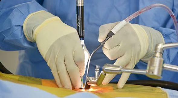
Endoscopic brain surgery is a minimally invasive technique in which the neurosurgeon uses a thin telescope with a camera and light (an endoscope) to reach and treat problems deep inside the head, either through the nose or through a small hole in the skull. For the right patient it can avoid a large skull opening, but it is not suitable for every tumour or condition, and some problems are still safer to treat with conventional surgery.
In Lahore, endoscopic techniques are most often used for pituitary tumours, certain skull base tumours, colloid cysts and hydrocephalus. This page explains what the operation involves, who may benefit and what recovery is usually like.
The endoscope gives a magnified, well-lit view on a screen, and fine instruments are passed alongside or through it. There are three main types:
Most pituitary tumours can be removed through the nose. This approach is described in detail on our endoscopic pituitary surgery page. Selected skull base tumours, such as some craniopharyngiomas, clival chordomas and tuberculum sellae meningiomas, may also be reached endonasally, and spinal fluid leaks from the skull base can be repaired the same way.
A colloid cyst is a benign cyst in the third ventricle that can block fluid flow and cause headaches or, rarely, sudden deterioration. Many symptomatic colloid cysts can be removed endoscopically through a burr hole.
When hydrocephalus is caused by a blockage (for example aqueductal stenosis or a tumour pressing on the fluid pathways), an ETV creates a small opening in the floor of the third ventricle so fluid can bypass the blockage. In suitable patients this may avoid the need for a permanent shunt.
The endoscope can also be used to biopsy tumours within the ventricles, open up fluid-filled cysts (such as arachnoid cysts) and assist during removal of some deep tumours as part of wider brain tumour surgery.
Suitability depends on the location, size and type of the problem rather than the patient's wish for a small incision. Endoscopic surgery may suit patients with:
It may not be the right choice when a tumour extends far to the side, wraps around major blood vessels or nerves, is very firm or very vascular, or lies on the surface of the brain where a standard craniotomy is straightforward. Very small nasal passages or active sinus infection can also affect the endonasal route.
Image guidance with neuronavigation is often used to confirm the safe route during surgery.
Most patients spend the first night in a high-dependency or intensive care area. Typical hospital stays are about two to five days, though this depends on the condition and any complications. Patients are usually helped to sit out and walk within a day or two.
After endonasal surgery, nasal blockage, crusting and mild headache are common for a few weeks. You will be asked not to blow your nose forcefully, strain or bend heavily for a period. Fluid intake and hormones are monitored after pituitary surgery. Many people return to desk work in about two to six weeks; physical work may take longer. Driving should wait until you are off strong painkillers and your doctor confirms it is safe, and longer if you have had seizures or visual problems. A follow-up MRI is usually arranged after a few months.
All brain surgery carries risks, and these are discussed individually. They include:
A craniotomy, where a section of bone is temporarily removed, may be safer when a tumour is large and extends sideways, is hard or very vascular, encases important arteries, or sits on the brain surface. Sometimes a combined approach is planned, or an endoscopic operation is converted to open surgery if bleeding or anatomy makes that the safer option. The aim is always the safest effective removal, not the smallest incision.
To find out whether your scan suits an endoscopic approach, you can see Dr. Khawar Anwar, a neurosurgeon in Lahore, by booking an appointment.
Usually not. The operation is done through the nostril, so there is normally no visible scar. Occasionally a small graft is taken from the thigh or abdomen to repair the skull base.
Not automatically. It can mean less brain retraction and a shorter stay for suitable lesions, but for some tumours a craniotomy is safer and allows more complete removal. The right choice depends on the scan.
For obstructive hydrocephalus in suitable patients, an ETV may avoid a shunt. It does not work for every type of hydrocephalus, and some patients still need a shunt later.
Nasal stuffiness and crusting usually improve over two to six weeks. Saline rinses and follow-up nasal cleaning may be advised.
That depends on the tumour type and whether any remains. Many benign pituitary tumours need only follow-up scans; others may need radiotherapy or medicines, decided after the tissue report.
Dr. Khawar Anwar is a Consultant Neurosurgeon & Spine Surgeon at PINS / Lahore General Hospital, Lahore, with over 10 years of experience in brain and spine surgery. He sees patients in Lahore and Gujranwala and focuses on minimally invasive and endoscopic techniques where they are suitable for the patient.
See the full list on the certifications page or read more about Dr. Khawar Anwar.
See all clinics, maps and timings on the locations page.
To book, call or WhatsApp 0333-7574732 and tell us which clinic and day suits you. Please bring your MRI or CT scan (films and CD), reports and a list of your current medicines to the consultation. For sudden weakness, loss of bladder or bowel control, a sudden severe headache or a head or spine injury, go to the nearest emergency department first.