
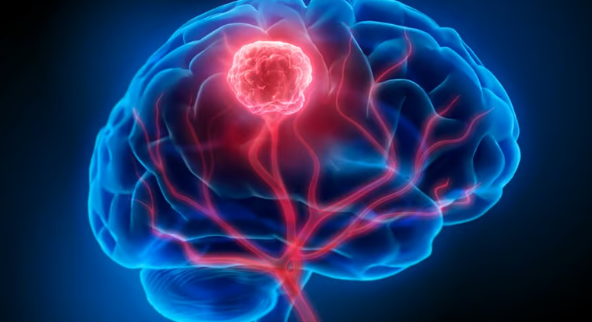
Brain tumour surgery aims to remove as much of a tumour as can be safely removed, relieve pressure on the brain and obtain tissue for an accurate diagnosis. Not every brain tumour needs an operation straight away: some small benign tumours are safely watched with scans, while others need surgery followed by radiotherapy or chemotherapy.
If you or a family member in Lahore has been told an MRI shows a "mass" or "lesion", this page explains the main tumour types, the symptoms to watch for, how the diagnosis is confirmed and what the different surgical options involve.
Brain tumours are either primary (starting in the brain or its coverings) or secondary/metastatic (spread from a cancer elsewhere, such as lung or breast). Primary tumours may be benign (non-cancerous, usually slow-growing) or malignant. Even a benign tumour can cause serious problems because the skull has no room to expand.
Usually benign growths of the pituitary gland that can affect hormones or press on the nerves of vision. Read about pituitary tumour surgery.
Mostly benign tumours arising from the membranes covering the brain. Small ones without symptoms are often monitored. See meningioma surgery.
Tumours arising from the brain's supporting cells, ranging from slow-growing low-grade gliomas to aggressive glioblastoma. Treatment usually combines surgery with oncology care. See glioma surgery.
Tumours at the floor of the skull near major nerves and arteries, such as acoustic neuromas, chordomas and craniopharyngiomas. See skull base tumour surgery.
Single or limited brain metastases may be removed surgically or treated with focused radiotherapy. Children may develop tumours such as medulloblastoma, which need specialist multidisciplinary care.
Symptoms depend on where the tumour is and how fast it grows:
Go to emergency if there is increasing drowsiness, repeated vomiting with headache, a prolonged seizure or sudden weakness. These may mean raised pressure inside the head.
The key test is an MRI of the brain with contrast (gadolinium), which shows the tumour's size, position, and how it relates to surrounding brain. CT is often done first in emergencies. Additional tests may include MR spectroscopy or perfusion, functional MRI for tumours near speech or movement areas, hormone tests and eye field testing for pituitary tumours, and body scans if a metastasis is suspected. Often, the final diagnosis is only made when tissue is examined under the microscope (histopathology).
The most common operation for brain tumours. A section of skull bone is temporarily lifted, the tumour is removed with a microscope, and the bone is replaced. Neuronavigation works like GPS to help plan the opening and locate the tumour. For tumours close to speech or movement areas, awake brain surgery may be considered, so brain function can be tested during the operation.
Pituitary tumours, some skull base tumours and tumours in the ventricles may be removed with an endoscope through the nose or a small burr hole. See endoscopic brain surgery for details and its limits.
When a tumour is deep, widespread or likely to respond to non-surgical treatment (for example lymphoma), a needle biopsy through a small hole may be the safest way to get a diagnosis before planning further treatment.
Small benign-looking tumours without symptoms, especially in older patients, may be monitored with repeat MRI scans, with surgery only if they grow or cause symptoms.
Surgery is often one step in treatment. Depending on the tissue report, radiotherapy, focused (stereotactic) radiosurgery or chemotherapy may be recommended. These are delivered by clinical oncology and radiation teams, and the plan is agreed together with the neurosurgeon. Some benign tumours need no further treatment beyond follow-up scans.
After a craniotomy, most patients spend a night in intensive care or high dependency and stay in hospital for around three to seven days, depending on the tumour and their condition. Headache and tiredness are common for several weeks. Steroids and anti-seizure medicines may be prescribed for a time. Patients with weakness or speech difficulty may need physiotherapy, speech therapy or occupational therapy. Return to work varies widely, from several weeks to a few months. Driving is usually restricted for a period, especially after seizures. Regular follow-up MRI scans are needed, sometimes for years.
Risks depend on the tumour's location and are explained before consent. They include bleeding, infection, brain swelling, seizures, new or worsened weakness, speech or vision problems, CSF leak, blood clots and anaesthetic risks. Some tumours cannot be completely removed safely, and regrowth can occur, which is why follow-up matters.
See a neurosurgeon as soon as a scan shows a brain lesion, or if you have symptoms such as new seizures or progressive headaches with neurological changes. At consultation, Dr. Khawar Anwar reviews the MRI with you, explains the likely type of tumour, whether surgery is needed now, and the options for treatment. Bring all scans, reports and a family member. You can book an appointment online.
No. Many brain tumours, such as most meningiomas and pituitary tumours, are benign. However, they can still cause symptoms by pressing on the brain and may need treatment.
Many benign tumours can be removed completely. For tumours that grow into important brain areas, the safest possible amount is removed, and the rest is treated with radiotherapy or monitored.
Usually only a small strip of hair along the incision line is shaved, and hair grows back over the following weeks.
That depends on the tumour and symptoms. Some need urgent surgery within days; others can be planned over weeks, and some only need monitoring.
Only if the tissue report and scan findings call for it. This decision is made with the oncology team after surgery.
Dr. Khawar Anwar is a Consultant Neurosurgeon & Spine Surgeon at PINS / Lahore General Hospital, Lahore, with over 10 years of experience in brain and spine surgery. He sees patients in Lahore and Gujranwala and focuses on minimally invasive and endoscopic techniques where they are suitable for the patient.
See the full list on the certifications page or read more about Dr. Khawar Anwar.
See all clinics, maps and timings on the locations page.
To book, call or WhatsApp 0333-7574732 and tell us which clinic and day suits you. Please bring your MRI or CT scan (films and CD), reports and a list of your current medicines to the consultation. For sudden weakness, loss of bladder or bowel control, a sudden severe headache or a head or spine injury, go to the nearest emergency department first.